We study microscale force sensing and optical imaging for biomedical analysis. Our research interests include microscale mechanical characterization, microfluidic analysis of cells, and biomedical microscopy/spectroscopy. Emphasis is on the study of micro/mesoscale tissues and cancer cells.
Micromechanical manipulation, imaging, and analysis of microtissues
Our approach relies on strong combinations of MEMS (microelectromechanical systems), robotics, optics, digital image analysis, and biological science.
We create unique robotic systems and analytical tools specifically designed for the study of micro/meso-scale (100 µm – 1 mm) tissues. The images below show a 4D digital image correlation (DIC) analysis of the micromechanical compression of a 3D organoid. Please also visit Microtissue Characterization and our recent publication for details. .
Using the tracked data, we can conduct a detailed structural analysis of volumetric deformation. The very important application of our method is the study of embryonic tissues. We found that the body of zebrafish embyo changes its stiffness rapidly as it grows. Details of the stiffness measurement are described in our paper. The growth of a zebrafish embryo can be seen in this video clip (1.0MB).
Please also read our related publications:
- Tomizawa et al. Journal of Biophotonics, 2022. link
- Tomizawa et al. Sensors 19(7), 1506, 2019. link
- Jaiswal et al. Biomedical Optics Express 2019. link
- Jaiswal et al. ACS Biomaterials Science & Engineering 2018. link
- Jaiswal et al. PLoS ONE 12, e0188346, 2017. link
Microfluidic and Lab-on-a-Chip systems
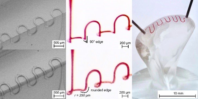
We have developed various microfluidic systems for cellular and tissue analysis. The image displays a 3D microfluidic channel fabricated using a custom 5-axis CNC milling machine. link
- Modarelli et al. Lab on a Chip, 25, 127-142, 2025. link
- Jaiswal et al. Journal of Magnetism and Magnetic Materials, 427, 7-13, 2017. link
- Chen et al. Scientific Reports, 5, 8745, 2015. link
- Chen et al. Lab on a Chip, 14, 446-458, 2014. link
- Huang et al. Biomedical Microdevices, 15, 673-681, 2013. link
- Hoshino et al. Analytical Chemistry, 84, 4292-4299, 2012. link
- Hoshino et al. Lab on a Chip 11, 3449-3457, 2011. link
Implantable sensors

We study an opt-mechanical flow sensor that minors the functionality of an implanted medical device.
- Garrett et al. Sensors and Actuators A, 312, 112110, 2020. link
- Soler et al. Yale Journal of Biology and Medicine, 91(3): 313–321, 2018. link
- Garrett et al. BMES 2019 Annual Meeting (BMES2019).
- M. Bao et al. The 43rd Annual Northeast Bioengineering Conference (NJIT 2017).
Biomedical microscopy/spectroscopy
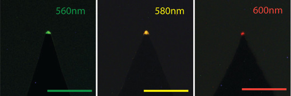
The techniques of optical microscopy and spectroscopy provide unique strength to our studies. Photograph shows an example of integrated quantum dot (QD)-based nanoscale LEDs integrated at the tip of near field scanning optical microscopy probe.
- Hoshino et al., Biomedical Optics Express, 5, 1610-1615, 2014. link
- Hoshino et al., Sensors and Actuators A, 216, 301-307, 2014. link
- Hoshino et al., Applied Physics Letters, 101, 043118, 2012. link
- Gopal et al., Applied Physics Letters, 96, 131109, 2010. link
- Gopal et al., Nanotechnology 20, 235201, 2009. link