Visco-elastography of 3D microtissue
We developed a new method of visco-elastography designed for the study of micro/mesocale tissues. We apply the elastography method to various light microscopy techniques, uniquely extending its capability beyond the commonly used ultrasound imaging or optical coherence tomography. See our YouTube video for the latest study, where we used light-sheet microscopy to characterize cell-by-cell viscoelasticity:
Also see our recent publication for details.
Micromechanical manipulation
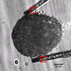
The main idea is to apply mechanical perturbation to the sample and analyze the resulting deformation through image analysis. One of the key tools we developed for micromechanical perturbation is the chopstick-like micromanipulator. It not only allows for the manipulation of micro/mesoscale tissues, but it is also capable of characterizing the mechanical properties the sample. We can measure the stiffness by studying bending of the cantilevers through image analysis. The images below show the stiffness analysis of a breast tumor spheroid and the von Mises strain mapping of a brain tumor spheroid. To our knowledge, our study is the first to quantify the elastic moduli of cancer spheroids! Read our publication for details.

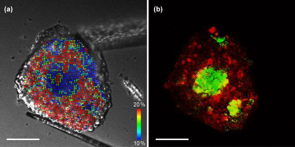
We conducted 3D imaging using differential interference contrast (DIC) and confocal microscopy. The figure above shows the analysis of a tumor-fibroblast co-culture spheroid. (a) Von Mises strain map calculated from DIC images. (b) Overlay confocal fluorescence of tumor cells (red) and fibroblast cells (green). We found solid fibroblast cells formed a core stiffer than surrounding cancer cells, which would not have been found only with conventional fluorescence images. Scale bars = 100 µm. See our publication for details.
A movie clip (link) is available from the article website.
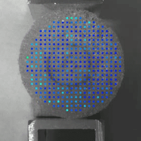
How it works: Image analysis of tissue deformation
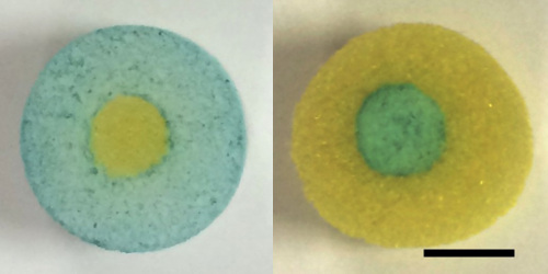
Here we explain the principle of image analysis-based strain (deformation) mapping. The photographs below show composite (core-shell) tissue phantoms made of two types of kitchen sponges (scale bar = 5 mm). One has the softer sponge in the core, and the other has the stiffer one in the core. Can you tell which is which? It is difficult just from still images.
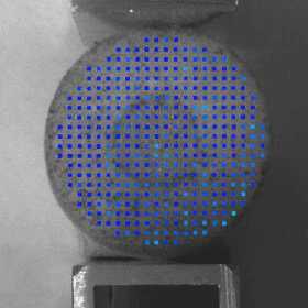
However, when you apply a deformation, you can see the difference. Softer materials deform more than stiffer materials, which can be observed as the amount of deformation distributed within the samples.
We used this technique to study the mechanical heterogeneity in silk fibroin protein-based, bioengineered biomaterials. Please read our publication for details.
Not only for research, now we use these tissue phantoms and the indentation system for K-12 education. See also Outreach Activities page for details.